Physiology 2130 Lecture Notes - Lecture 6: Inhibitory Postsynaptic Potential, Astrocyte, Prolactin

29 May 2018
School
Department
Course
Professor

Module 6 pt 1 – Nervous System
Intro:
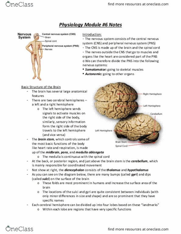
• The nervous system consists of the central nervous system (CNS) and peripheral nervous system (PNS)
o The CNS is made up of the brain and spinal cord
o The PNS is made up of the nerves outside the CNS that go to muscles and organs
• The PNS can be further divided into
o somatomotor – going to skeletal muscles
o autonomic – going to other organs
Basic Structure of the Brain:
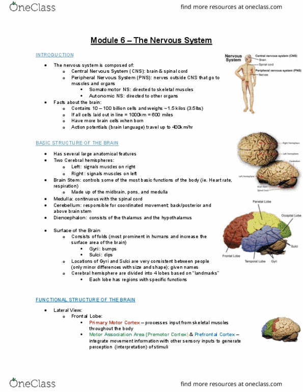
• There are two cerebral hemispheres
o The left hemisphere sends signals to activate muscles on the right side of
the body, and vice versa
• The brain stem
o Controls some of the most basic functions of the body
▪ Like: HR, respiration, etc.
o The brain stem is made up of the midbrain, pons and medulla oblongata
▪ The medulla is continuous with the spinal cord
• At the back or posterior region and just above the brain stem is the cerebellum
o Responsible for coordinated movement
• The diencephalon is made up of the thalamus and hypothalamus
• There are many bumps (gyri) and dips (sulci) on the surface of the brain
o These folds are most prominent in humans and increase the SA of the brain
o The locations of the gyri and sulci are quite consistent across individuals and
are so prominent they have different names
• Each cerebral hemisphere can be divided into four lobes
o Parietal, occipital, frontal and temporal
Functional Structure of the Brain:
• Each region of the brain has a specific function, and some are organized in an extremely specific manner
• Note: four different views of the brain, different structure from each view
• Lateral view
o Frontal lobe:
▪ Primary cortex: processes input from skeletal muscles throughout the body
▪ Premotor cortex (motor association area) and the prefrontal cortex: integrate
movement information with other sensory inputs to generate perception of stimuli
o Parietal lobe:
▪ Primary somatosensory cortex: receives input from the major sense organs
▪ Association areas: integrate sensory information with other association area of the
cortex to for meaningful perceptions
o Cerebellum:
▪ Processes sensory information and coordinated the execution of movement in the
body
▪ As the structure with the largest number of neurons in the brain, the cerebellum
receives input from somatic receptors, receptors fore equilibrium and balance and
motor neurons from the cortex
o Temporal lobe:
▪ Primary auditory cortex and auditory association areas: receive and process signals
from auditory nerves and integrate them with other sensory inputs
▪ Other areas: involved in olfaction and mediating short-term memory storage and
recall
find more resources at oneclass.com
find more resources at oneclass.com

o Occipital lobe:
▪ Responsible for vision
▪ Primary visual cortex: receives input directly from the optic nerve
▪ Visual association areas: further process visual information and integrate it with
other sensory inputs
• Medial view
o Corpus collosum:
▪ Dense bundle of nerve fibers that serves as a pathway and connection between the two cerebral
hemispheres
▪ This connection allows the brain to integrate sensory and motor information from
both sides of the body and coordinate whole-body movement and function
o Pituitary gland:
▪ Primarily regulates endocrine organs
▪ The anterior pituitary is derived from epithelial tissue of the pharynx
▪ The posterior pituitary derives from neural tissue of the hypothalamus
▪ Anterior pituitary hormones – LH, FSH, ACTH, TSH, GH and prolactin
▪ Posterior pituitary hormones – vasopressin and oxytocin
▪ Pituitary function is regulated by the hypothalamus
o Pons:
▪ Acts as a relay station for transferring information between the cerebellum and the
cerebral cortex
▪ Also coordinates and controls breathing
o Diencephalon:
▪ Thalamus: receives sensory inputs from the spinal cord and integrates sensory
information before sending it to the cortex
▪ Hypothalamus: controls a variety of endocrine functions, mainly through directing
the release of hormones
o Midbrain:
▪ Or mesencephalon
▪ Bridges the lowers brainstem with the diencephalon
▪ Primary function is controlling eye movements and exerts some control over
auditory and visual motor reflexes
o Medulla
▪ Portion of the brainstem that has primary control over involuntary functions such as
breathing, blood pressure and swallowing
▪ Fibres from the corticospinal tract, which originate in the motor cortex, cross over
to the opposite side of the spinal cord to innervate muscles on the opposite side of
the body
• Ventral view
o Optic chiasma:
▪ Optic nerves from each eye meet here, where they cross over and continue on to
the lateral geniculate bodies on the thalamus
▪ From there the axons extend to their respective hemisphere on the primary visual area of the
occipital lobe
o Brain stem:
▪ Extension of the spinal cord and consists of three regions
▪ From left to right on figure
• Midbrain, pons and medulla
▪ The brainstem is the center for many involuntary functions
▪ Incorporates 9 cranial nerves
• Dorsal view
o Primary motor cortex
▪ Posterior end of the frontal lobe
▪ Processes information relating to skeletal muscle movement
find more resources at oneclass.com
find more resources at oneclass.com
Document Summary
The pns can be further divided into somatomotor going to skeletal muscles: autonomic going to other organs. There are two cerebral hemispheres: the left hemisphere sends signals to activate muscles on the right side of the body, and vice versa. The brain stem: controls some of the most basic functions of the body. The diencephalon is made up of the thalamus and hypothalamus. Functional structure of the brain: each region of the brain has a specific function, and some are organized in an extremely specific manner, note: four different views of the brain, different structure from each view. From there the axons extend to their respective hemisphere on the primary visual area of the occipital lobe: brain stem, extension of the spinal cord and consists of three regions. From left to right on figure: midbrain, pons and medulla, the brainstem is the center for many involuntary functions. Serves as a general interpretative center for visual and auditory information.