Physiology 3120 Lecture Notes - Lecture 8: Homeostasis, Vasoconstriction, Baroreflex

9 May 2018
School
Department
Course
Professor

Physiology 3120
Dr. Stavraky
Lecture 8
Lecture Outline
Blood Vessel Structure
- How their structure relates to their function
Cardiovascular Regulatory Mechanisms
- 3 types of regulation
1. local control mechanisms
2. humoral regulation
3. neural control mechanisms
o baroreceptor reflex
Blood Vessel Structure
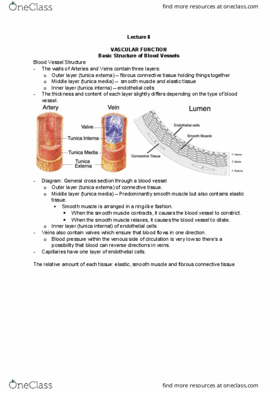
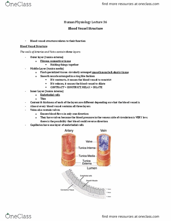
- The walls of Arteries and Veins contain three layers:
o Outer layer (tunica externa)
▪ fibrous connective tissue
o Middle Layer (tunica media)
▪ Find specialized tissue like smooth muscle (arranged in ring) (smooth muscle
contraction or relaxation causes vessel to constrict or dilate) and elastic tissue
o Inner Layer (tunica interna)
▪ endothelial cells
- the thickness and contents of each of these layers is different depending on what vessel it is
- Veins also contain valves
- Capillaries have one layer of endothelial cells
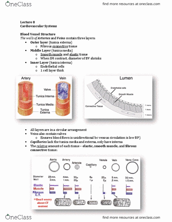
- The relative amount of each tissue: elastic, smooth muscle and fibrous connective tissue
- Don’t need to memorize all these different numbers – just get a general idea of their relative
sizes and relative thicknesses
- Aorta
o very large vessel (size of a loonie) but its wall is very thin
o contains a lot of elastic tissue
o there is some smooth muscle and some fibrous connective tissue to hold it together
- arteriole
o very thick wall relative to their size
o contain a lot of smooth muscle to regulate the diameter of the vessel
- capillary
o very thin bc only 1 cell layer thick
o doesn’t contain any elastic or smooth muscle or fibrous connective tissue
find more resources at oneclass.com
find more resources at oneclass.com

- vena cava
o larger diameter than aorta
o wall thickness is even thinner than aorta
o contains some smooth muscle
o recall: one way to increase venous return is to activate SYN to contract the smooth
muscle in the walls to send more blood back to heart (increase venous return) to
increase EDV to increase SV
- how does the blood vessel structure relate to its function?
Structure relates to Function
- tie together the previous diagram to the pressure curve
- in the aorta and arteries, the blood pressure is very pulsatile – bc the blood contracts and sends
the blood to the vessels and these vessels are going to expand
- when the ventricles contract during systole, they eject a certain amount of blood (even at rest,
they are ejecting 70mL of blood)
- that blood is going to enter the aorta and arteries and bc they are thin walled with lots of elastin
tissue they can expand to accommodate that in rush of blood – they are very compliant
- when the heart is relaxing, they contract and continue to push the blood along
- bc they can expand, there is very little resistance (very little drop in BP) and so the large arteries
are very good at distributing the blood along the body this is exactly what they are – our
distribution vessels
- so we maintain a high BP through these vessels and we can distribute the blood throughout the
body
- when we get to the arterioles, we have the largest drop in BP bc it’s the site of highest resistance
- there is a lot of resistance here bc they are very small and bc they have a very thick wall with
very little elastin tissue in it so they are not going to expand like the arteries do
- so the arterioles are called the resistance vessels
- but they do have a lot of smooth muscle which allows the vessels to relax for vasodilation or
contract for vasoconstriction to allow us to alter blood flow
- when we get to our capillaries, we have the largest total cross sectional area and the slowest
blood velocity
- this is perfect bc those two combined with a very short diffusion distance makes our capillaries
our exchange vessels (where exchange takes place)
- when we get to our veins, bc this is where we find most of our blood at rest (70% of it), and they
have larger diameter shows that they can accommodate more blood
- they also have thin walls and they are easy to expand bc they contain elastin vessels
- our veins are called out capacitance vessels
find more resources at oneclass.com
find more resources at oneclass.com
Document Summary
How their structure relates to their function. 3 types of regulation: humoral regulation, neural control mechanisms, baroreceptor reflex. Inner layer (tunica interna: endothelial cells the thickness and contents of each of these layers is different depending on what vessel it is. Capillaries have one layer of endothelial cells. The relative amount of each tissue: elastic, smooth muscle and fibrous connective tissue. Don"t need to memorize all these different numbers just get a general idea of their relative sizes and relative thicknesses. Structure relates to function tie together the previous diagram to the pressure curve the blood to the vessels and these vessels are going to expand. Most tissues have the capacity to control their own blood flow by a process called autoregulation a process by which individual vascular beds maintain a relatively constant blood flow when moderate changes occur in blood pressure. Also, when a blood vessel constricts, pressure increases upstream and decreases downstream.