PSL300H1 Lecture Notes - Receptive Field

Document Summary
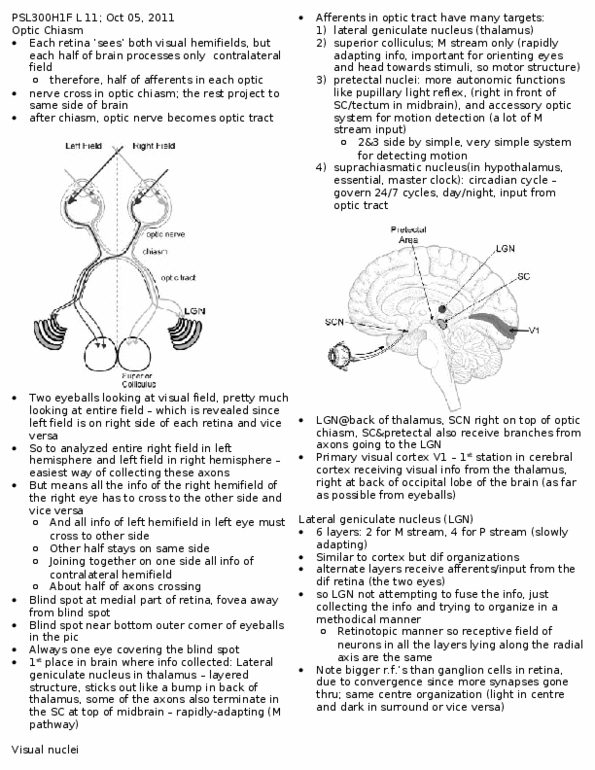
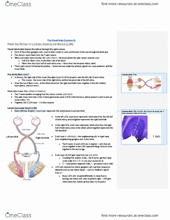
All visual information (light/dark contrast) is conveyed by ganglion cells, which are the only cells with axons that produce action potentials that project to the brain. Lateral geniculate nucleus (lgn) back of thalamus ~ visual nucleus receives information from both m and p streams (but terminate in different layers) important for visual perception sends signals to primary visual cortex. Lgn aligns all receptive fields so cells match up information is sent up to the visual cortex where fusion occurs have center-surround receptive fields, but bigger receptive fields than ganglion due to convergence. V1 lies along calcarine sulcus in medial wall of occipital lobe at back. Lgn organizes inputs to visual cortex of brain retinotopic organization o layout on retina is preserved in lgn and v1 largest area devoted to fovea and macula, where photoreceptors are most numerous (small receptive fields, more packed in)