[Kinesiology 3337A/B] - Final Exam Guide - Everything you need to know! (41 pages long)

30 Mar 2017
School
Department
Course
Professor
Document Summary
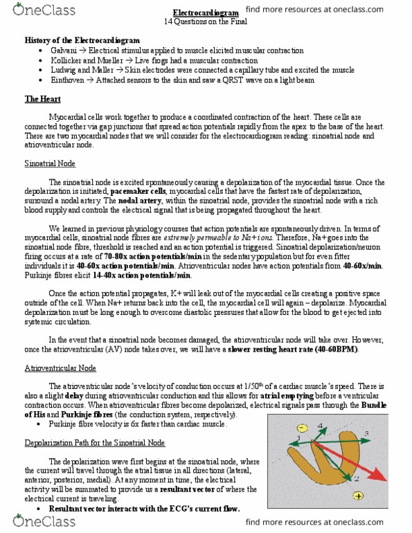
Galvani electrical stimulus applied to muscle elicited muscular contraction. Kollicker and mueller live frogs had a muscular contraction. Ludwig and maller skin electrodes were connected a capillary tube and excited the muscle. Einthoven attached sensors to the skin and saw a qrst wave on a light beam. Myocardial cells work together to produce a coordinated contraction of the heart. These cells are connected together via gap junctions that spread action potentials rapidly from the apex to the base of the heart. There are two myocardial nodes that we will consider for the electrocardiogram reading: sinoatrial node and atrioventricular node. The sinoatrial node is excited spontaneously causing a depolarization of the myocardial tissue. Once the depolarization is initiated, pacemaker cells, myocardial cells that have the fastest rate of depolarization, surround a nodal artery. The nodal artery, within the sinoatrial node, provides the sinoatrial node with a rich blood supply and controls the electrical signal that is being propagated throughout the heart.