PS267 Chapter Notes - Chapter 4-5: Parietal Lobe, Visual Cortex, Temporal Lobe

Document Summary
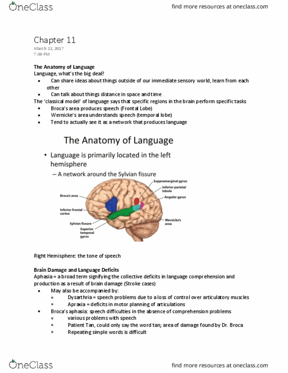
Split brain research: way to study the specializations of each hemisphere by presenting tasks to only one hemisphere (right vs left visual field) The 2 hemispheres do not represent information in the same way. Macroscopic (what can we see visually without microscrope) anatomical asymmetries not lined up perfectly. Right hemisphere has more volume in the frontal region. Left hemisphere has more volume in the occipital region. Planum temporale: wernicke"s area, larger in left, language comprehension. Look at differences in circuitry in homotropic areas; language is the most obvious to tell. Wernicke"s area in the left has more space between cortical columns (broca"s area is the left too but more to the front) Cell sizes are larger in the left hemisphere. Work together in large part because of corpus callosum: largest white matter structure in the brain that connect the left and right hemisphere. Commissures: smaller bands of fibers connecting the two hemispheres. Anterior commisure: primarily connects some temporal lobe regions.