ECON 546 Lecture Notes - Lecture 10: Glucocorticoid Receptor, Sonication, Methyl Group

27 Jun 2018
School
Department
Course
Professor

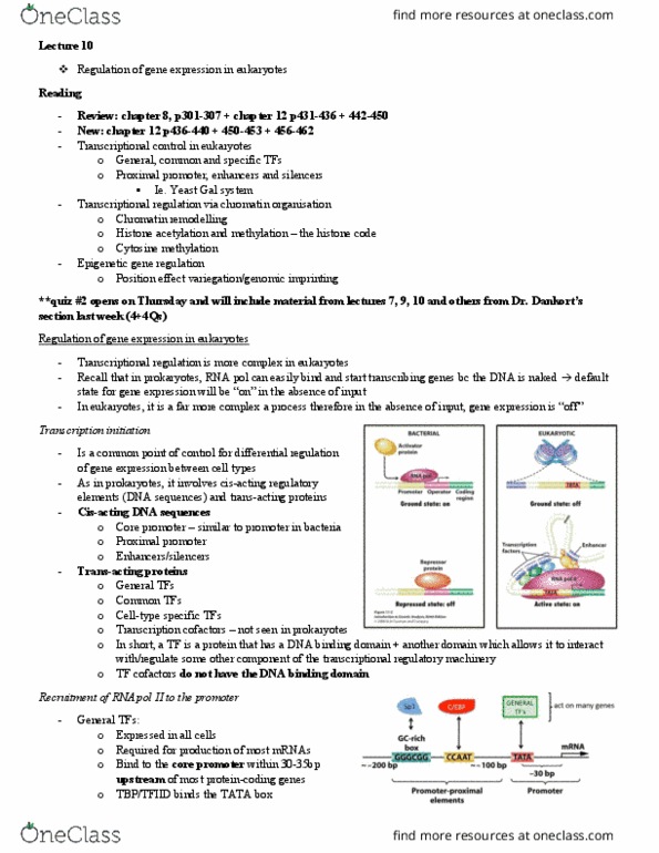
BIOL 568 – Najafabadi L9: 6/2/18
L9: Current approaches to study gene regulation
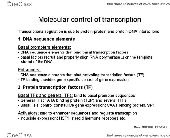
Recap: transcription factors
oDNA-binding protein that regulate transcription. Can bind to promoter-proximal elements or
enhancers.
oHow TFs recognise their targets – DNA sequences ‘motifs’
Modelling the sequence binding preferences of TFs:
oE.g. 8 known binding sequences for TF (ROX1) in yeast. All similar
oConsensus binding site: summary of all the different types of sequences that the TF might
bind to. E.g. almost always has C in second position etc.
oY and H – examples of IUPAC representation of ambiguity
oN = any base
oPosition-specific frequency matrix (PSFM or PFM): using numbers – better than IUPAC
representation as it shows the actual number of times each specific base is observed at a
particular position.
oMotif logo: produced from the PSFM. Visual representation – height of each letter is
proportional to number of times that nt is seen in that position.
oCalculating probabilities: 6As and 2 Ts at position 1. Probabilities are 0.75 and 0.25,
respectively. Can then calculate the probability of any particular sequence.
oBetter than counting as counts depends on the number of observations.
Probabilities are independent of the number of the observed binding sites –
normalised.
oUse probability matrix to make the visual motif
oDifferent representations of PSFM: First binding position doesn’t matter too much – first
column is scaled down, as TF will generally still bind. The middle of sequence is most
important
Is the probability of seeing the motif in a GCN4 binding site the same as probability of it binding to
the sequence?
oNo. Many things affect the probability of the binding of GCN4 e.g. binding affinity to that
particular sequence, concentration of GCN4, whether it is accessible or not (e.g.
heterochromatin vs. euchromatin).
Where do PSFMs come from? A brief introduction to in vitro methods
SELEX: systematic evolution of ligands by exponential enrichment
oRandom pool of oligonucleotides (synthesised in vitro).
oImmobilized protein (TF) used to select oligonucleotides. Some happen to have binding
sequence by chance.
oBound and unbound then separated
oRepeat step by amplification of bound sequences – selecting for sequences that bind to the
TF,
1
find more resources at oneclass.com
find more resources at oneclass.com

BIOL 568 – Najafabadi L9: 6/2/18
oRepeat until each TF has at least one bound.
oThen identify the sequence to reveal the binding site. As number of cycles increases, there is
more and more of the binding site.
oAlign sequences so that similar parts of the sequences are on top of each other. Then count
and produce PSFM.
PBM: protein-binding microarrays
oSurface with hundreds of thousands of different oligonucleotides attached of known DNA
sequence. Create ds strand from ss using DNA Pol.
oAdd labelled protein e.g. fluorescent antibody. Wash away unbound protein.
oVisualise under microscope – see spots where protein binds = DNA molecule has the binding
site
oAlign
Where to find PSFMs?
Databases of TF binding models:
oCIS-BP: data collected from all types of experiments – two strands, reverse complement
Different combinations of TFs binding – orientation affects regulation
oJASPA: curated set of TF motifs
oUniPROBE: PBM motifs
In vivo identification of TF binding sites: a brief introduction to ChIP-seq
ChIP-seq: chromatin immunoprecipitation sequencing
oPrefer to do in vivo – what really happens inside the cell. Many factors inside the cell that
cannot be reproduced inside the test-tube e.g. heterochromatin; cooperativity of TFs – need
cofactors that may not be known to be provided.
oTF bound to DNA.
oTake chromatin sample and fragment (e.g. by sonication)
oSeparate fragments that are bound by the TF using an antibody.
oImmunoprecipation – pulling out a protein from a solution
oSeparate and purify the DNA from the TF, then sequence. Small
fragments then mapped to genome. Some fragments map to the
positive strand, others to the negative strand.
Sequences that bind to positive strand pile up on LHS of TF
binding and negative strand on the RHS.
Length of DNA ~50-100bp
Fragmentation gives a much longer fragment but only one end
is sequenced ~50bp. Hence the curve. Actual binding site is
2
find more resources at oneclass.com
find more resources at oneclass.com
Document Summary
Recap: transcription factors: dna-binding protein that regulate transcription. Can bind to promoter-proximal elements or enhancers: how tfs recognise their targets dna sequences motifs". Modelling the sequence binding preferences of tfs: e. g. 8 known binding sequences for tf (rox1) in yeast. All similar: consensus binding site: summary of all the different types of sequences that the tf might bind to. Visual representation height of each letter is proportional to number of times that nt is seen in that position: calculating probabilities: 6as and 2 ts at position 1. Can then calculate the probability of any particular sequence: better than counting as counts depends on the number of observations. Is the probability of seeing the motif in a gcn4 binding site the same as probability of it binding to the sequence: no. Many things affect the probability of the binding of gcn4 e. g. binding affinity to that particular sequence, concentration of gcn4, whether it is accessible or not (e. g. heterochromatin vs. euchromatin).