MGY277H1 Lecture Notes - Lecture 2: Periplasm, Fluorescence Microscope, Lac Repressor

30 Mar 2018
School
Department
Course
Professor
Document Summary
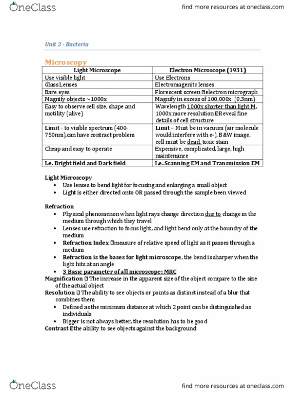
Unit 2 the bacteria: magnification: increase in apparent size of object (e. g. 4x appears 4 times larger) Directs light towards specimen at an angle, only light scattered by specimen enters objective. Cells stand out bright against dark background (think about how laser can"t be seen from side unless there"s smoke) Expensive, complicated, large can see up to visible spectrum (400-750 nm) wavelength 1000x shorter than light, 1000x. Easy to see cell size, shape, motility more resolution (0. 3 nm) Must be in vacuum, images in black and white, see more details, cell must be dead. Electrons scanned over surface, released electrons are reflected and observed. Stains purple in gram stain (retain dye, decolorizing agent thought to dehydrate the thick peptidoglycan layer which then acts as barrier against dye from leaving) Stains pink in gram stain (decolouring agent damages outer membrane, thin layer of peptidoglycan cannot retain dye) Thin peptidoglycan layer between two membranes (space between called periplasm)